
Uterine fibroids · Focused ultrasound
An incision-free treatment option for fibroids: HIFU
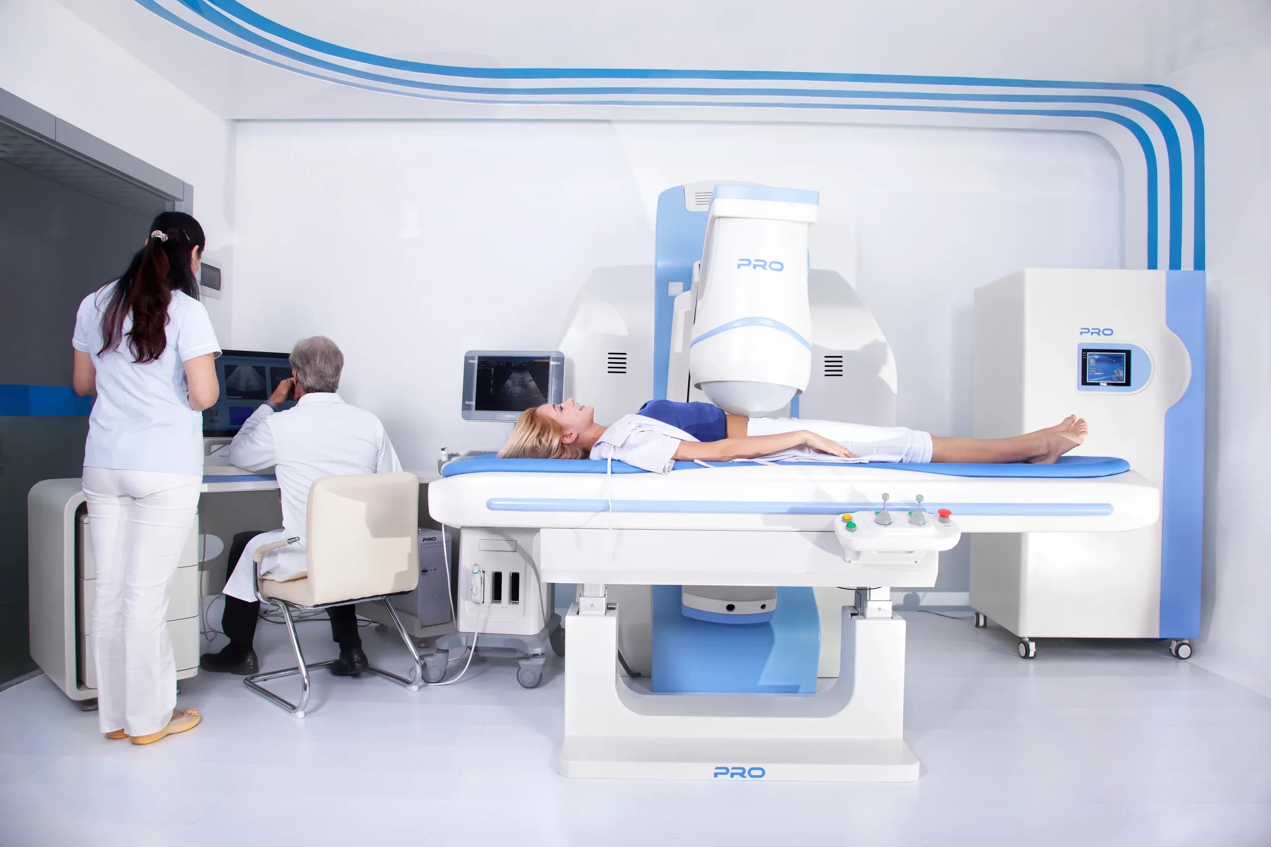
Ultrasound energy is delivered from a probe outside the body and focused on a single point inside tissue; no skin incision is needed. It is not applicable to every fibroid or every patient — eligibility is determined with imaging.
- No incision, no stitches
Energy is delivered from a probe outside the body.
- Imaging-guided
The procedure advances under real-time ultrasound.
- Eligibility is assessed with MRI
MRI findings and clinical history are read together.
How does HIFU work?
- 01
Planning
The target volume and the beam path are defined on imaging.
- 02
Focusing
Energy is emitted from the probe outside the body and converges at a single point.
- 03
Real-time monitoring
The procedure advances step by step under real-time ultrasound guidance.
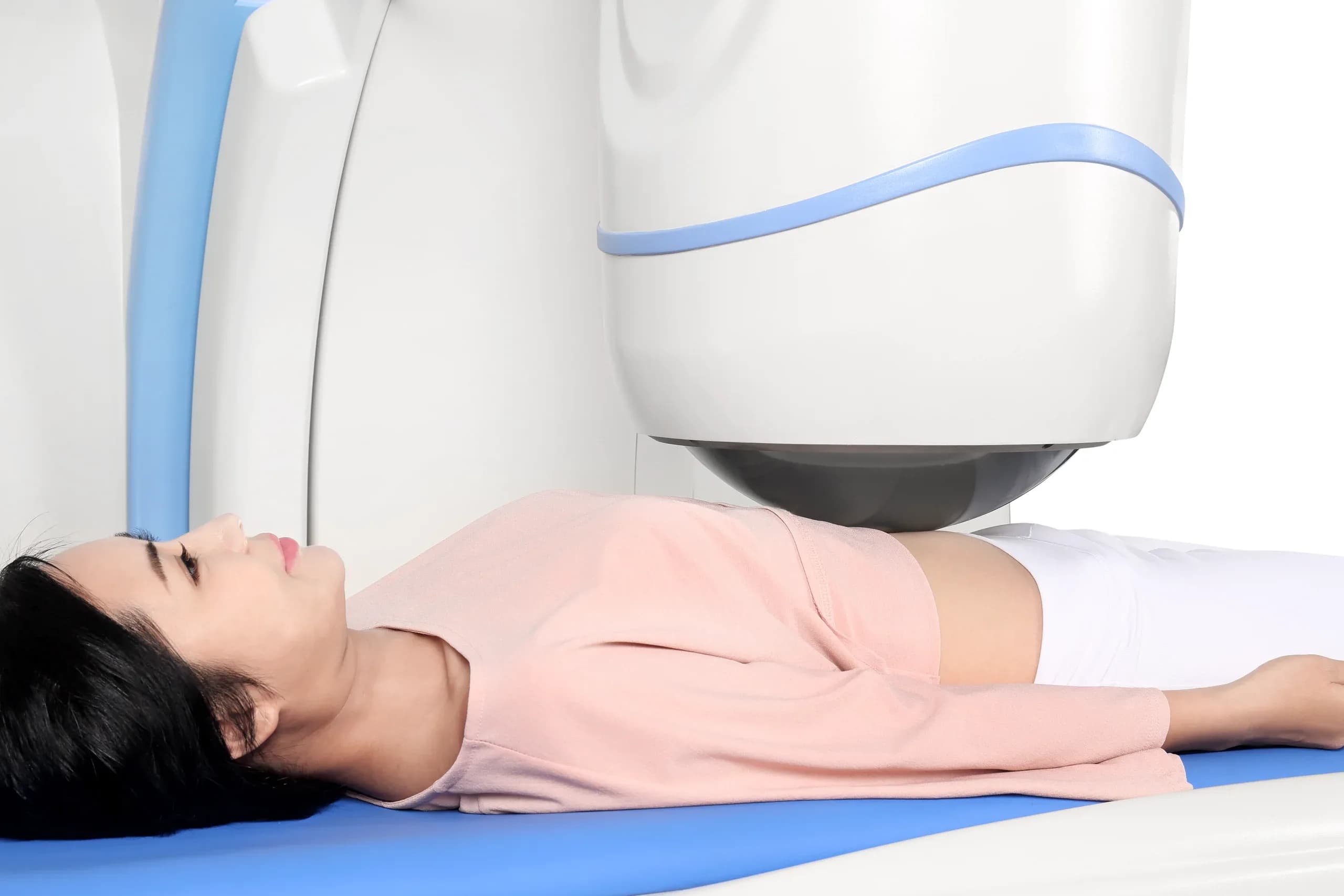
The treatment probe sits outside the body, above the abdominal wall; a water bolus carries the sound into tissue. The energy stays diffuse along the path and concentrates only where it is focused.
Bowel segments or scar tissue in the beam path are among the situations that limit applicability.
An honest frame
What HIFU offers — and what it does not promise
Describing a method honestly means stating what it does and where it stops, on the same page.
What stands out about HIFU
- No incision or stitches
Energy comes from outside the body; no surgical incision is made.
- The uterus is not removed
The method targets fibroid tissue; it does not require removing the uterus.
- Performed under imaging guidance
Target volume and beam path are planned on imaging and monitored throughout.
- Recovery is its own topic
Clinical series report a short return to daily life; the duration varies by person and by the extent of the procedure.
It is not suitable in every case
- Fibroid location
Position within the uterine wall directly affects applicability.
- Size and number
Assessing one fibroid is not the same as assessing many.
- Technical suitability
The beam path and the fibroid’s imaging characteristics decide whether targeting is possible.
- Clinical history and priorities
Pregnancy plans, comorbidities and previous treatments all enter the assessment.
This list is not an eligibility test; which heading is decisive becomes clear only with imaging.
Process
Could HIFU be suitable for me?
No online test can answer that. Eligibility is determined by reading the headings below together with your imaging.
What the assessment looks at
Location
Submucosal, intramural or subserosal position changes both symptoms and method choice.
Size
Size alone is not a threshold; the same diameter means different things in different positions.
Number
The number of fibroids changes the volume to be planned and the process.
Symptoms
Bleeding, pressure, pain or no complaint at all — the first heading that decides whether treatment is needed.
Clinical context and priorities
Age, pregnancy plans, comorbidities and the person’s own priority.
How the assessment works
Gathering the information
A short form lets you send the basic information a physician needs for a preliminary review, in an organised way.
Physician’s preliminary review
Your MR images, symptoms, previous treatments, pregnancy plans and general health are considered together.
Getting back to you, and the consultation
You are contacted through the channel you prefer; the definitive assessment is made in consultation with a physician.
These three steps are the part verified today. Treatment planning and the steps that follow are settled by physician assessment.
What is on the table?
These approaches are not rivals; they are different answers for different situations.
Lower procedural burden
- Surveillance
- Medical management
- HIFU
- Embolisation
- Myomectomy
Higher procedural burden
Surveillance
When there are no symptoms or only mild ones, monitoring at set intervals is an option in its own right.
Medical management
Medication may aim to bring symptoms — bleeding in particular — under control.
Myomectomy
Surgical removal of the fibroid; the uterus is preserved and the technique varies with the fibroid's location.
Embolisation
A vascular procedure in which the vessels feeding the fibroid are blocked with the aim of shrinking it.
HIFU
The focus of this site
Ultrasound energy focused from outside the body targets fibroid tissue without an incision.
Two systems, one method
The technology behind the treatment
The devices are not a product showcase but the technical layer of how the method is delivered. Both apply ultrasound-guided focused ultrasound; they differ in how the treatment probe is carried.
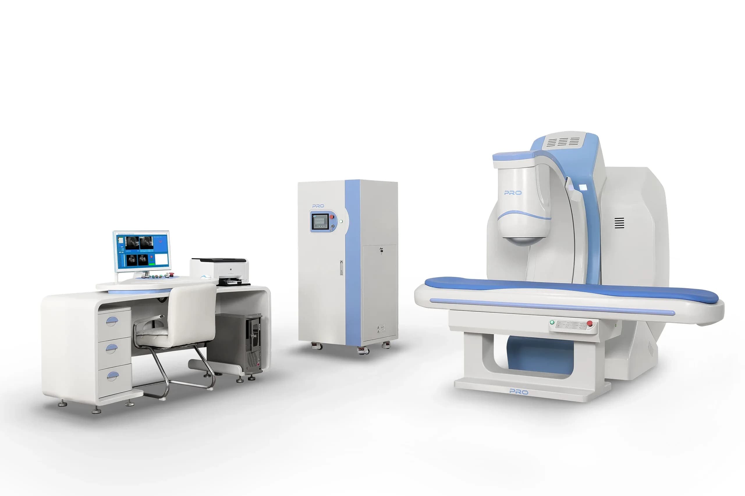
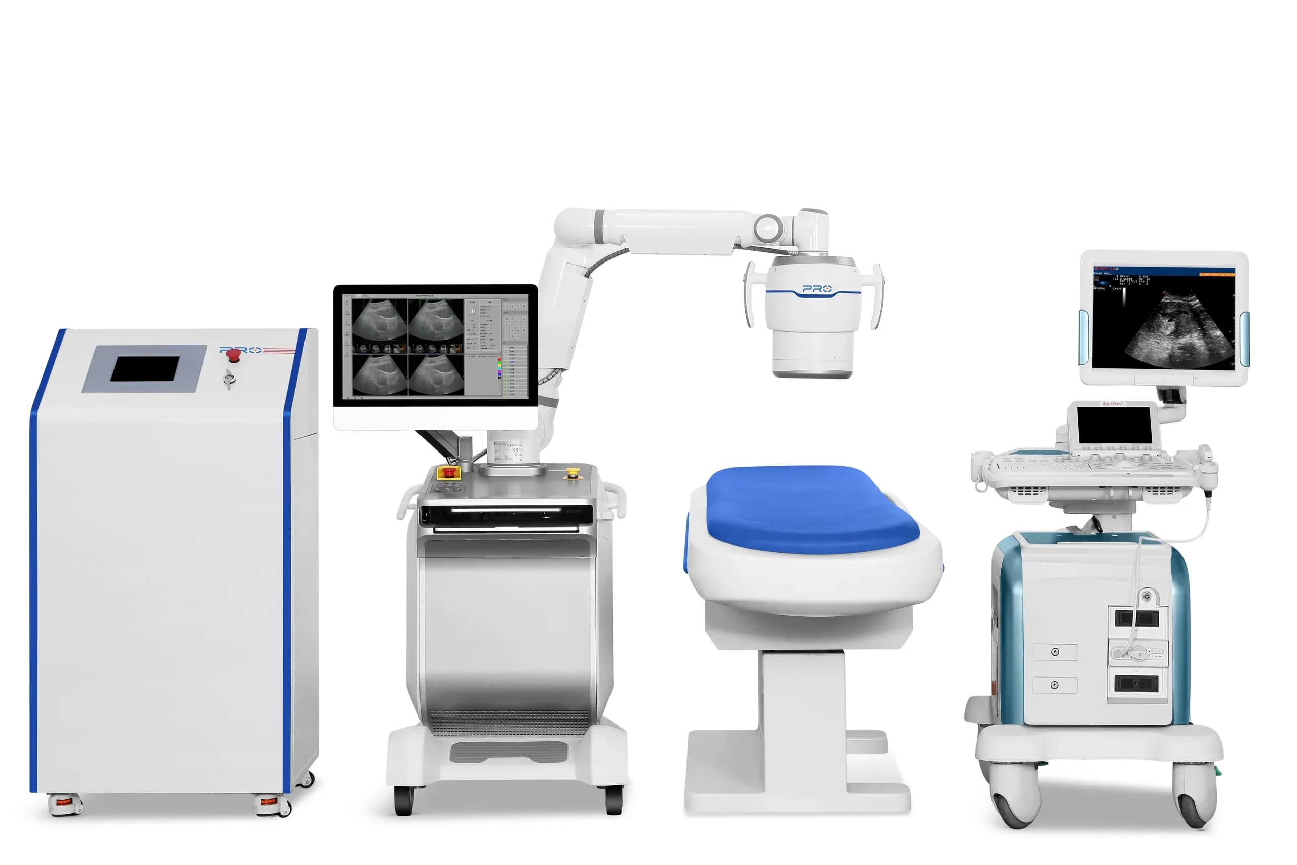
From manufacturer documentation — not a photograph of our centre · Technical details come from the manufacturer’s official documentation; they carry no claim of clinical effectiveness.
The systems are brought into Türkiye through the authorised distributor Asel Tıp Medikal, the legal counterparty for import, installation and technical service. Both systems are listed as “Registered” in the TİTCK product tracking system.
Team
Who carries out the assessment?
Clinical content is reviewed by a physician; the assessment and the treatment decision are made by a physician.
Evidence
What does the evidence say, and where is it limited?
Systematic reviews and meta-analyses exist on HIFU for fibroids; every critical claim on these pages is listed with its citation.
The limit: a significant share of the literature is single-centre series and long-term data is still limited. That is why we do not present a study’s result as “your result”.
34 publications in the bibliography · most recent 2026
What do evidence levels A / B / C mean?
- A
- Statements resting on several peer-reviewed publications or reviews, listed with their sources on the page.
- B
- Statements resting on a limited number of publications or single-centre series, listed with their sources.
- C
- Technical information from manufacturer documentation or regulatory records; not evidence of clinical effectiveness.
- D
- General information and process description that does not require a citation.
From the bibliography
- Meta-analysisReproductive outcomes of high-intensity focused ultrasound ablation and myomectomy for uterine fibroids: a systematic review and meta-analysis
- Meta-analysisLong-term outcome and risk factors of reintervention after HIFU ablation for uterine fibroids: a systematic review and meta-analysis
- Meta-analysisReproductive and Obstetric Outcomes after UAE, HIFU, and TFA of Uterine Fibroids: Systematic Review and Meta-Analysis
Frequently asked questions
How many HIFU sessions are needed?
For uterine fibroids, HIFU is usually planned as a single session; the duration depends on the size and number of fibroids. In some cases an additional session or a different method may be needed to reach the intended ablation volume — this is assessed at the planning stage.
Is the HIFU procedure painful?
Heat and pain sensations can occur during treatment, which is why the procedure is typically performed with sedation and pain control while you remain able to communicate. Mild pain for a few days afterwards is commonly reported.
Does HIFU remove the fibroid completely?
HIFU aims to inactivate fibroid tissue with heat (ablation); the tissue is not surgically removed but shrinks over time as the body resorbs it. The amount of shrinkage varies with the patient and the extent of ablation, and some patients may need further treatment.
Who is not a candidate for HIFU?
Pregnancy, suspected malignancy and certain fibroid types (such as pedunculated fibroids) are common exclusion criteria in clinical studies. Situations where the ultrasound beam cannot reach the target safely may also rule out treatment. The final decision always requires individual assessment.
Why is an MRI needed before HIFU?
MRI shows the location, number and tissue characteristics of your fibroids — features that directly determine whether HIFU is technically feasible and likely to be effective. That is why imaging sits at the centre of the eligibility assessment.
Can fibroids grow back after HIFU?
Fibroids that receive sufficient ablation usually shrink and stop causing symptoms; however, regrowth of a treated fibroid or development of new fibroids is possible. The likelihood of retreatment is closely linked to patient selection and the extent of ablation achieved.
Next step
Let’s start the assessment
A short form lets you send the information a physician needs for a preliminary review.
The form does not generate an eligibility result or medical advice.